Microscope Endodontics
Dental operating microscopes offer high magnification and fiber optic illumination that reveal details inside of the tooth that are invisible to the naked eye. This allows the dentists to see each tooth’s unique anatomy, enabling precise treatment such as locating hidden or calcified canals or diagnosing cracks or fractures. Microscopes are essential in enhancing the quality of endodontic care and in supporting improved outcomes.
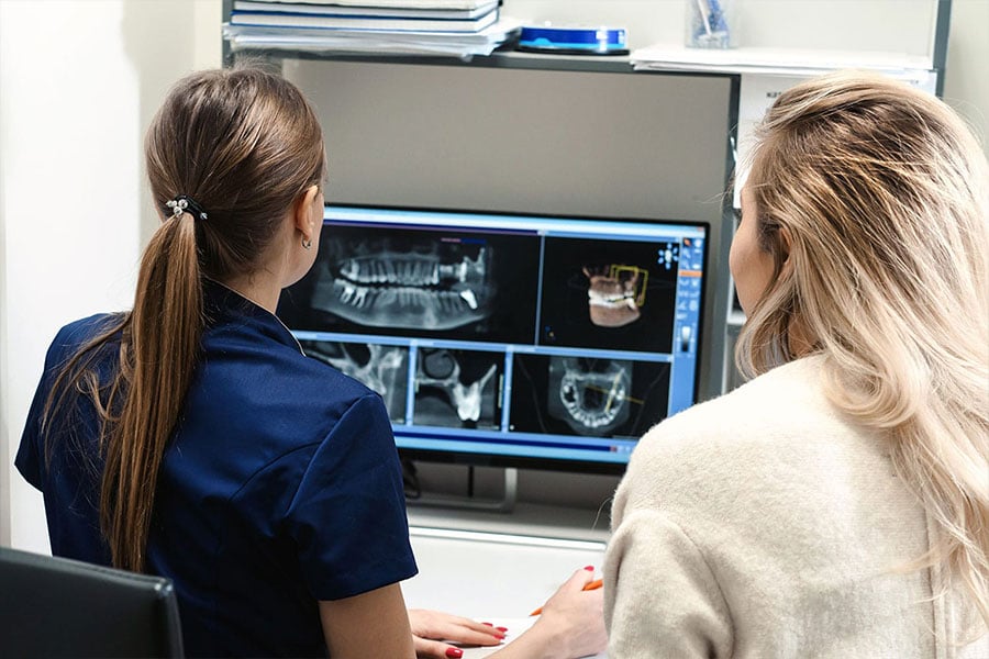
Cone Beam CT Scanner
Our on-site 3D CBCT provides crystal-clear, three-dimensional views. This revolutionary tool helps our doctors accurately assess tooth anatomy, sinuses, nerves, and other vital landmarks before procedures, ensuring optimal treatment planning.
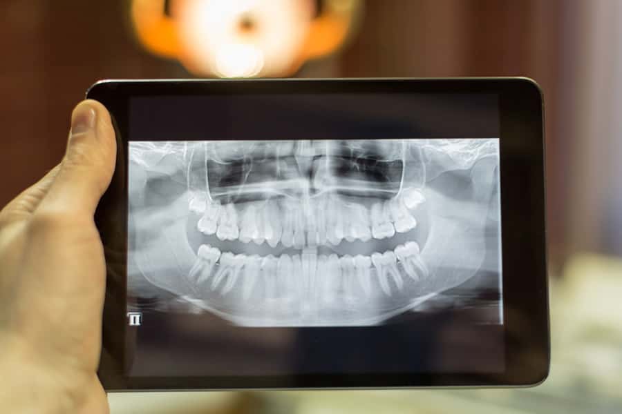
Digital Radiography
Digital x-rays capture high-quality images with significantly less radiation than traditional methods. The images are instantly available and can be viewed on large screens in each treatment room. Your endodontist will review the images with you and provide a personalized assessment, including a review of your diagnosis and the recommended treatment options. Your doctor will answer any questions you have before starting any procedure.
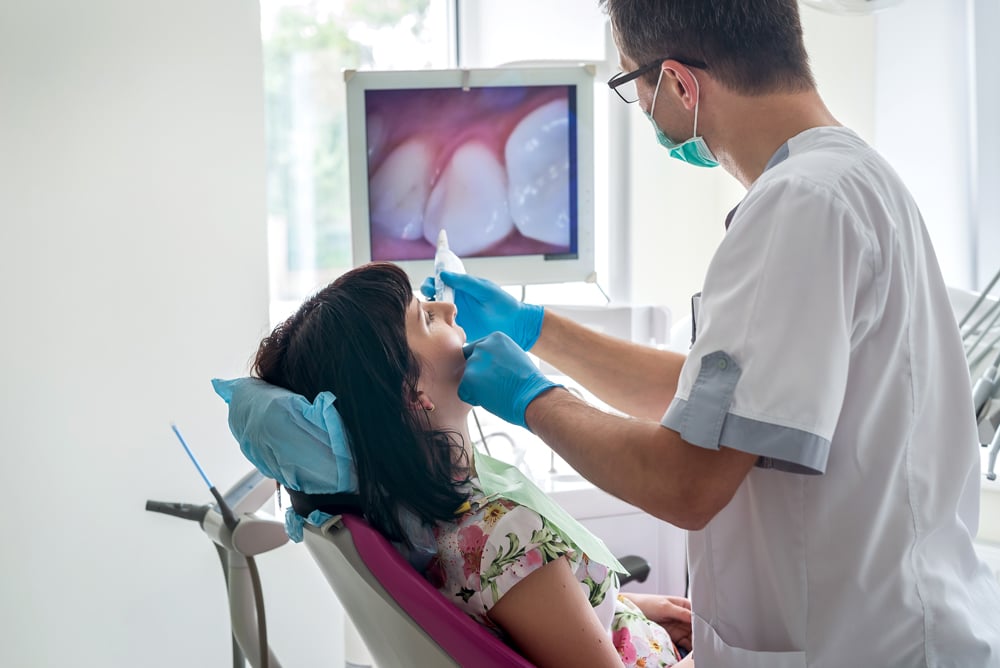
Intra-Oral Camera
Intra-oral cameras capture images of what the doctor is seeing through the microscope, helping to communicate details about your treatment and document findings. This tool will enhance your understanding of your treatment and support shared decision-making with your dentist.